Skeletal
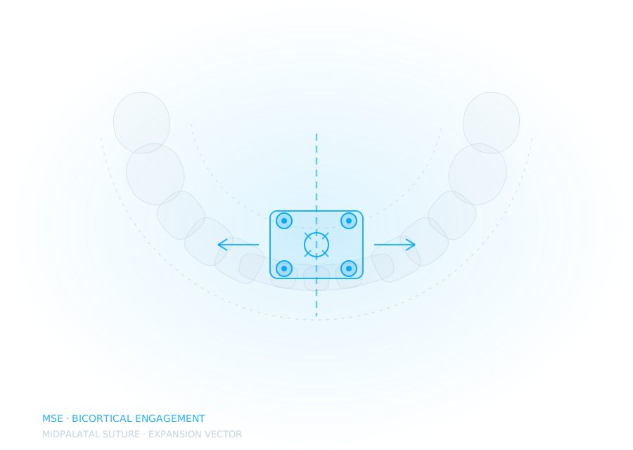
MSE in a 24-year-old, suture opened
Bicortical screw placement planned on CBCT after the first plan showed insufficient palatal bone posteriorly. Screws moved 4mm anterior; suture split at day nine.

Every case records what we planned, what changed, and what we would do differently. The one we turned down is in here too, because that decision was the useful part.
Worked examples of how each case type is planned and documented. The illustrations are schematic representations of the workflow, not clinical records.
Bicortical screw placement planned on CBCT after the first plan showed insufficient palatal bone posteriorly. Screws moved 4mm anterior; suture split at day nine.
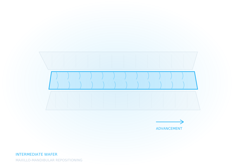
Le Fort I with BSSO. Intermediate wafer indexed to 1.5mm so it located reliably without binding under drapes.
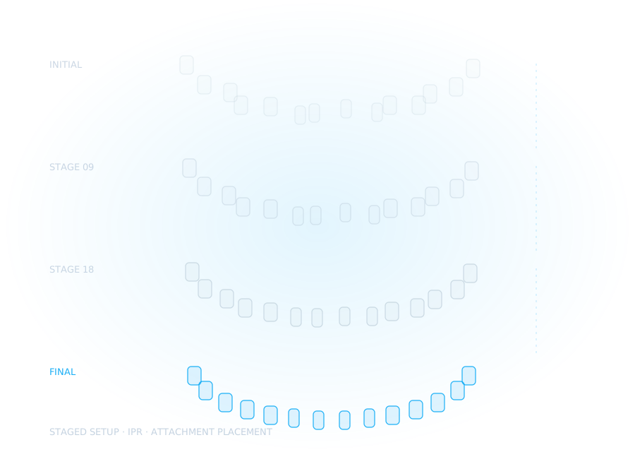
Staged with IPR distributed across three phases rather than front-loaded, so anchorage held without a TAD.
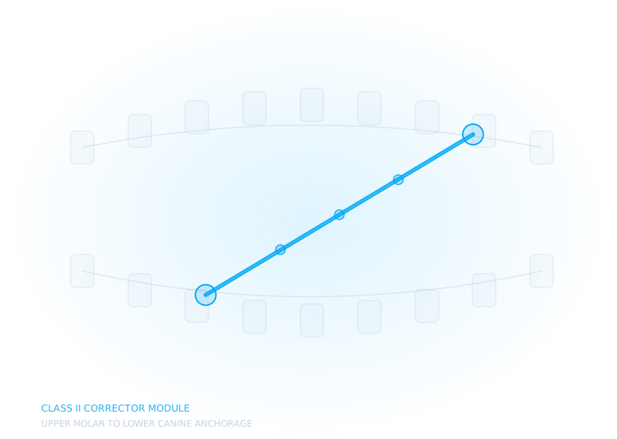
Construction bite recorded at the chair; anchorage assessment flagged the lower incisors and a lingual arch was added before fitting.
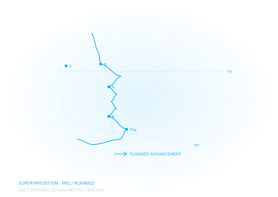
Superimposition of the surgical plan against the twelve-month record. The chin projection landed 2mm short of plan and we documented why.
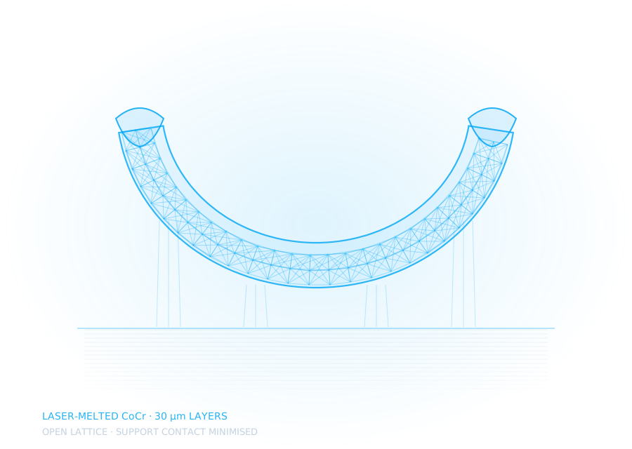
Replaced a soldered-band design that had fractured twice at the same junction. Printed as a single body, so there was no joint left to fail.
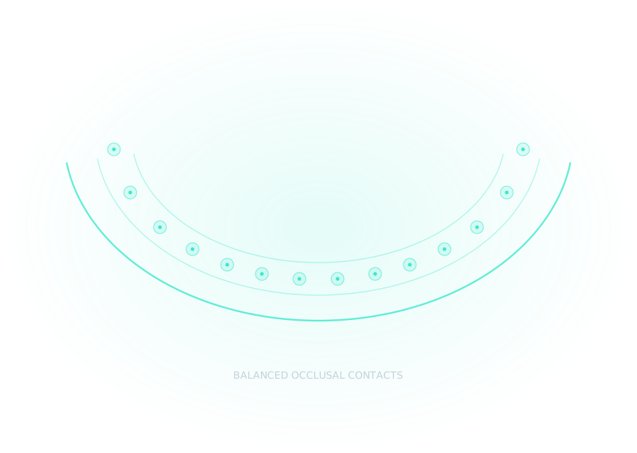
Designed at a defined vertical with even bilateral contacts, printed in clear resin and worn for eight weeks before planning.
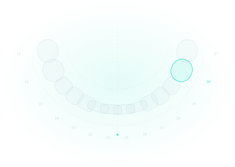
Planning showed cortical thickness below the threshold at every viable screw site. We said no and suggested a surgically assisted approach instead.
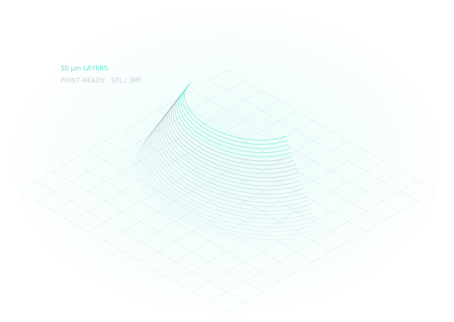
Nested and oriented for the practice's own printer, with base thickness and support contact tuned to their resin.
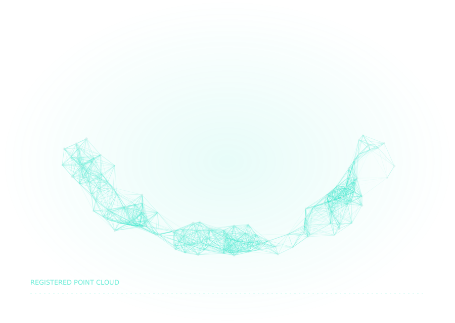
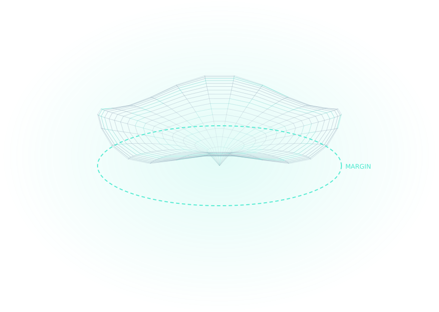
The original scan stopped at the gingival margin, so there was no vault to build a frame on. We specified the re-scan and the case ran clean.
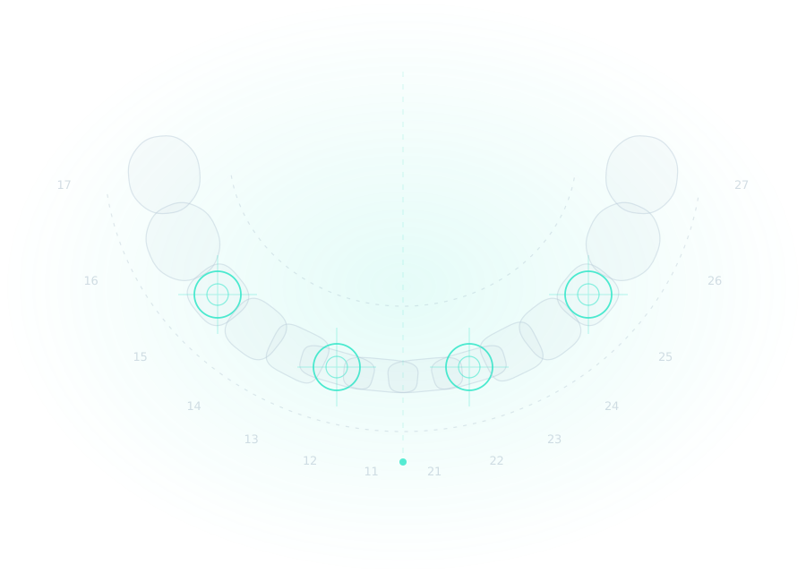
Bone-driven position would have put the screw access through the buccal cusp. Replanned from the restoration back; angle changed 14 degrees.
Emergence built from the healing abutment scan, with screw access rotated out of the central fossa before the design was approved.
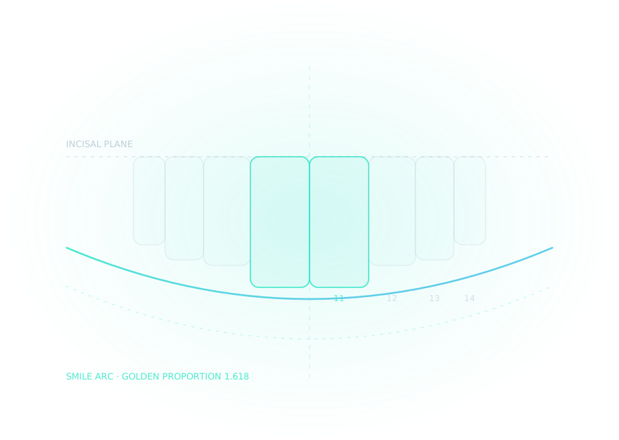
Space opening at 12 and 22 set by the final restoration width, not by whatever the aligners happened to produce. The prosthesis drove the tooth movement.
An expander that looked perfect on screen and loosened at week three is a design that was wrong. We only learn that if someone writes down what happened in the mouth.
Nothing goes into this library without documented patient consent for the images used, and identifying details are removed as a matter of course. Orthodontic records are unusually identifying — a profile photograph and a ceph together are close to a name. Treat them accordingly.
This is general guidance, not legal advice. Check your own regulator’s rules on advertising and patient imagery.
Send us something difficult. We will design it, document the reasoning, and you keep the write-up either way.