
What we need from you
- CBCT covering the full palate, the midpalatal suture along its length, and the nasal floor. A field that clips the posterior palate cannot be planned on
- Intraoral scan of the upper arch, including the vault to its full depth — not stopped at the gingival margin
- Age and skeletal maturity. Chronological age is a weak proxy; a hand-wrist or cervical vertebral assessment is better if you have one
- Photographs, and the transverse discrepancy you are trying to correct
The three questions we answer before designing anything
1. Will the suture open?
Midpalatal suture maturation is stageable on CBCT — the Angelieri classification is the common reference — and the later stages, where interdigitation and bony bridging are established, are where non-surgical expansion starts failing. In a 24-year-old this is the question, not a formality. We stage it and tell you what we see.
If the suture reads as fused, that is not a design problem to be engineered around. It is a different treatment plan, and we will say so.
2. Is there bone for the screws?
Palatal bone thickness is not uniform. It is generally most generous anteriorly and paramedian, and thins posteriorly — which is inconvenient, because posterior placement is where the expansion force is often most useful. We measure at each candidate site and report the number.
Where the posterior sites will not support bicortical engagement, the honest options are to move the screws anteriorly and accept the altered force distribution, or to change approach. Both are better than placing a screw into bone that cannot hold it and discovering the fact under load.
3. Where do the screws go without hitting anything?
Root proximity, the incisive canal and the nasal floor all constrain the usable window. Planning on the volume rather than on the cast is the entire point of doing this digitally.
Bicortical, and why it matters
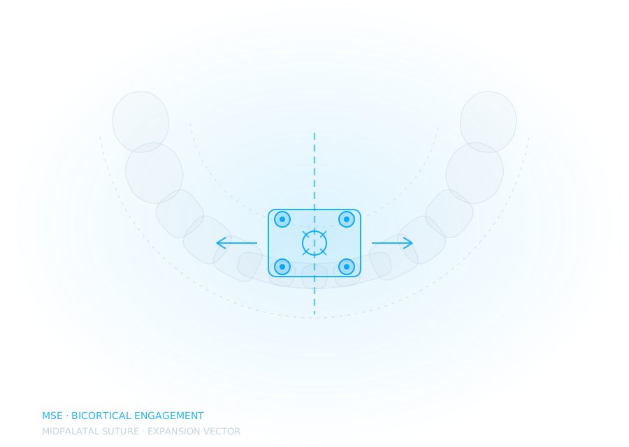
Engaging both the palatal and nasal cortices gives the screw markedly better stability under the forces expansion generates. Monocortical placement works, but it is working harder, and screw loosening under load is the failure mode that quietly turns a skeletal expansion into a dental one.
We plan for bicortical where the anatomy allows it and tell you where it does not, site by site, rather than reporting an average.
What leaves the lab
- The framework design, with the expander body positioned for the force vector the plan needs
- Screw positions with measured bone thickness at each, and whether each is bicortical
- An insertion guide, so the planned position is the placed position
- Band or bonded pad design to suit the abutment teeth
- The suture maturation assessment, in writing
Where these cases go wrong
- Screws placed into insufficient bone — loosening under load, and the expansion becomes dental
- A suture that was never going to open, planned as though it would
- Asymmetric opening, usually traceable to asymmetric anchorage rather than to the appliance
- Framework flex, which absorbs the activation instead of transmitting it
Background on choosing between the approaches: MSE, MARPE and SARPE — what decides which.
Timeline
Three to four working days from a usable CBCT and scan, including the assessment. If the CBCT will not support planning you will hear that on day one, not day four.
Planning and design support only. Diagnosis, treatment planning and the clinical decision remain entirely with the treating clinician.