
A wafer that does not seat is rarely a wafer problem. It is a record problem, or a registration problem, that stayed invisible until the one moment it could not be ignored. By then the surgeon is holding it, the anaesthetist is watching the clock, and someone has to decide whether to force it, trim it, or abandon it.
What follows is the order worth checking — starting with what can still be salvaged in theatre and working back to the things that were decided weeks earlier.
1. The condyles are not where they were when you took the bite
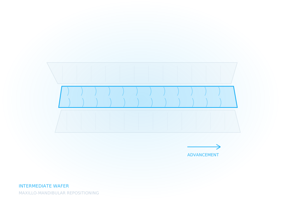
The intermediate wafer positions the maxilla against the unoperated mandible. Every millimetre of its accuracy therefore depends on the mandible being in the same position in theatre as it was when the record was taken. Under general anaesthesia, with the muscles fully relaxed, the condyles seat differently from how they sit in a conscious patient who is being asked to bite.
If the planning record was taken in a habitual or protruded position rather than a properly seated one, the wafer will be built to a mandible that no longer exists once the patient is asleep. It will look correct on screen and bind in the mouth.
This is the one on the list that is genuinely difficult to detect beforehand, because the design is faithful to the record it was given. The design cannot know the record was wrong.
2. The dentition was misregistered onto the CBCT
The wafer's fitting surface comes from the intraoral scan; its spatial position comes from the CBCT. Those two datasets have to be merged, and the merge is only as good as the surfaces used to align it.
Where it goes wrong:
- Scatter from restorations, brackets or bands degrades the dental surfaces in the volume, precisely the surfaces the registration algorithm is trying to match
- Too few registration points, or points clustered in one region, so the merge is accurate anteriorly and drifts posteriorly
- Motion artefact during the scan, which does not always announce itself but distorts the geometry the merge relies on
A registration error of a fraction of a millimetre at the incisors becomes considerably more at the molars. The wafer seats anteriorly and rocks, which is the pattern that most often points here.
3. The indexing is too deep for the tissue on the day
Indexing has to be deep enough to locate the arch unambiguously and shallow enough to seat over tissue that is not in its planned state. In theatre, with drapes limiting access and soft tissue starting to respond to surgery, a wafer that indexed beautifully on the model can bind on the very undercuts that made it feel secure.
This is the failure that is usually recoverable at the chair — selective relief of the binding point, if you can identify it quickly. It is also the one most easily prevented at design stage by deciding indexing depth deliberately rather than accepting a default.
4. The teeth moved between planning and surgery
Pre-surgical orthodontics does not stop because a scan was taken. If there is a gap of weeks between the records and the operating date, and active mechanics are still running, the wafer is built to an arch that no longer exists.
Wire changes, elastics and continued levelling all move the surfaces the wafer sits on. This one is entirely predictable and almost entirely preventable. It is a scheduling problem wearing a technical costume.
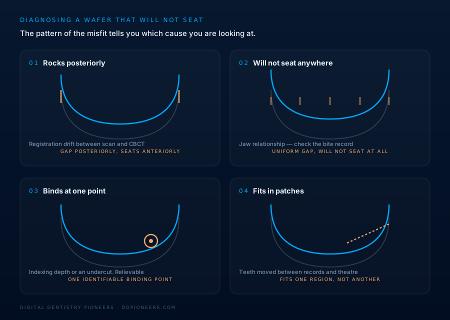
How to tell which one you are looking at
The pattern of the misfit narrows it quickly:
- Seats anteriorly, rocks posteriorly — look at the registration first
- Will not seat at all, everywhere — suspect the jaw relationship, particularly on an intermediate wafer
- Seats but will not go fully home, one identifiable point — indexing depth or an undercut; relievable
- Fits some teeth and not others, in a region, the arch has changed since the record
Which is most likely
Ranked by how difficult they are to catch before theatre rather than by raw frequency, the order above is deliberate. The condylar position problem heads the list because it is the only one that is invisible to everyone downstream, the records look complete, the merge succeeds, the design is faithful, and the error is baked into the reference the whole plan was built on.
Registration error is second because it is detectable, but only if someone looks for it specifically. Indexing depth is third and is largely a design decision. Dental movement between records and surgery is last not because it is rare but because it is entirely a scheduling matter, and everyone involved already knows the dates.
Put differently: the further up this list a cause sits, the fewer people had a chance to catch it.
What we do about it
Every surgical case here includes a planning session before a wafer is designed. Not a form, a conversation about the movement, the record, and how the bite was taken. It exists because three of the four causes above are decided long before anyone opens a CAD file, and none of them can be designed out afterwards.
We also report what we measured, so if a wafer does not seat you can work backwards through the plan instead of guessing.
Planning support and design only. Diagnosis, treatment planning and the surgical decision remain entirely with the treating clinician and surgeon.