
A design is a set of decisions made about anatomy. If the record of that anatomy is wrong, incomplete or ambiguous, the decisions are guesses wearing a serial number, and no amount of care at the CAD stage recovers it.
These five account for the overwhelming majority of what we send back. Each takes under two minutes to prevent.
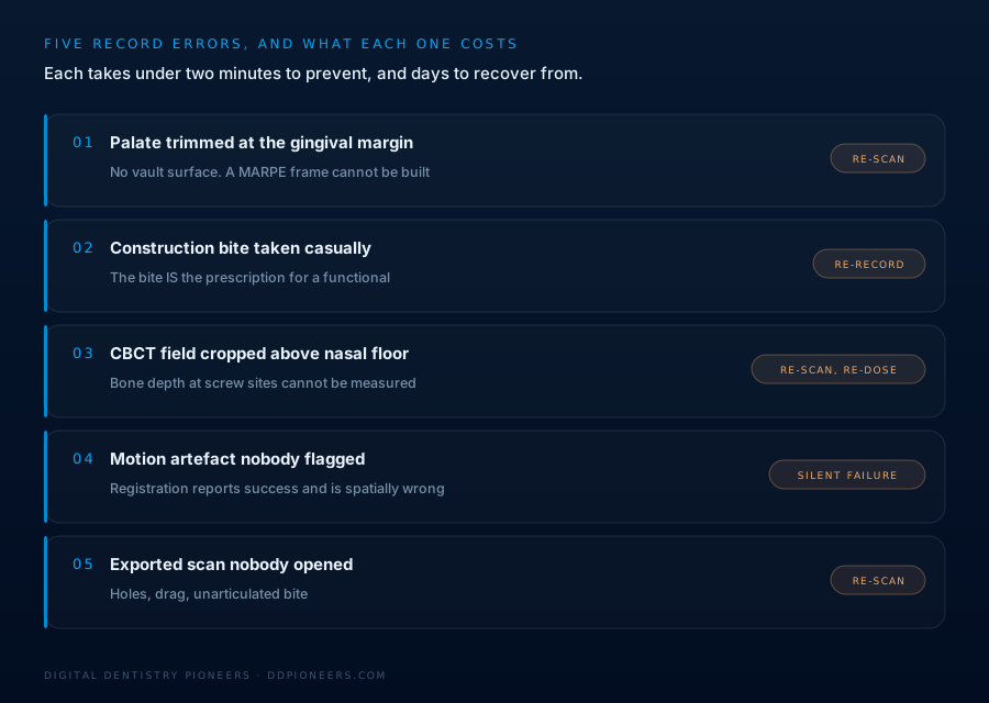
1. The palate trimmed at the gingival margin
Most scanners default to a trim that keeps the teeth and a collar of gingiva. That is perfectly adequate for a crown and useless for an expander.
A MARPE or MSE frame is built on the palatal vault. The framework spans it, the miniscrews go through it, and the clearance between the appliance body and the mucosa is calculated from it. If the scan stops at the gingival margin there is simply no surface there to design against.
What we can do with a trimmed palate is guess the vault contour from the arch form. What we will not do is guess and then hand you a frame that either impinges on tissue or stands so far off it that the anchorage is compromised.
Prevention: scan the full vault as a deliberate step, not as an accident of the trim setting. Sweep the palate slowly, most scanners struggle on a smooth, featureless surface, and rushing it produces exactly the holes you will be asked to re-take.
2. The construction bite taken casually
For a functional appliance, the construction bite is not a record of where the patient is. It is a prescription for where you want them to end up. It determines advancement, vertical opening and the midline the appliance will hold.
Taken quickly, with the patient posturing unevenly or the vertical guessed, it produces an appliance that is technically well made and clinically wrong. And it will be wrong in a way that only becomes visible weeks later.
Prevention: record it deliberately, note the vertical you used and why, and tell us. "Construction bite, 6 mm advancement, 4 mm vertical at the incisors, midline coincident" takes ten seconds to write and removes all ambiguity.
3. The CBCT field cropped above the nasal floor
Reduced field of view is good practice, less dose, and often better resolution. But the field has to contain the anatomy the plan depends on.
For skeletal expansion, that means the midpalatal suture along its length, the nasal floor, and enough vertical extent to read palatal bone depth at the intended miniscrew sites. A volume cropped above the nasal floor cannot support any of those measurements.
The frustrating version of this is when the field is technically adequate but the region of interest sits at the very edge, where reconstruction artefact is worst. The data is present and not trustworthy.
Prevention: when ordering the scan, say what it is for. A radiographer setting a field for "orthodontic assessment" makes different choices than one setting it for "miniscrew-assisted palatal expansion planning".
4. Motion artefact nobody flagged
This is the quiet one. A patient who moved slightly during acquisition produces doubling, blurring or stepped edges. On a quick scroll through the axial slices it can look acceptable, but the surfaces the merge algorithm relies on are precisely the ones degraded.
The result is a registration that reports as successful and is spatially wrong. Every measurement afterwards inherits that error, and nothing downstream reveals it. The first sign is often an appliance that does not fit and no obvious reason why.
Prevention: check the coronal and sagittal reconstructions, not just the axials. Look at the cortical outlines. Motion shows as a double line or a shear step. If you see it, re-take. A repeat scan costs less than a re-designed appliance and a delayed patient.
5. The scan nobody looked at
The most common of the five and the easiest to fix: the scan was exported and sent without anyone opening it afterwards.
Holes where the software interpolated. Soft tissue dragged into the occlusal surface. A cheek retractor captured as anatomy. Two arches that do not articulate because the bite scan caught the patient mid-slide. All visible in about fifteen seconds of rotating the model.
Prevention: open the exported file, the actual export, not the scanner's preview, and rotate it once before sending. That is the whole procedure.
The two-minute check
Before anything is exported, in this order:
- Rotate the model and look underneath it. Holes and soft-tissue drag are obvious from below and invisible from the occlusal view the scanner shows you
- Confirm the arches articulate. Open the bite scan and check the teeth meet where you expect, not where the software assumed
- On any skeletal case, look at the palate specifically. Not the teeth, the vault
- On any CBCT, scroll the coronal and sagittal reconstructions. Motion shows as a double cortical line or a step. Thirty seconds
- Write the objective in one sentence before you send. If you cannot state it in a sentence, the design will not be able to either
What happens at our end
We check records before designing on them, and we flag problems the same day rather than the day before you needed the appliance. If something is borderline we will tell you it is borderline and let you decide, rather than quietly designing around it.
If you are not sure whether a set of records will support the case, send them and ask. We would much rather answer that question than start the clock on a design we will have to abandon.
Planning and design support only. Diagnosis, treatment planning and clinical decisions remain entirely with the treating clinician.